Assign the level on CT or MR, and know how much to trust it — imaging tier first, then the ranked fallback landmarks, then Castellvi typing of a transitional vertebra.
One reliable foundation: the only definitive way to number lumbar vertebrae is to count caudally from C2 on whole-spine imaging. Every landmark shortcut is a fallback with quantifiable error. When landmarks disagree, or a transitional vertebra is present, get whole-spine imaging before any level-targeted procedure. In the body reading room this is nearly always CT — the topogram usually reaches the cervicothoracic junction and sagittal and coronal reformats are already on the study, so the tools that settle the count are the ones you have.
Step 1
What are you looking at?
This sets the ceiling on confidence before any landmark is used. The question is how far cephalad you can see — a topogram or MR localizer is not the same as coverage of the ribs, and neither is the same as whole-spine imaging.
Before settling for a lower tier: a chest radiograph, or the topogram of any prior CT in the record, lets you count ribs for free. Spine radiographs in the record are frequently never separately reported — if they are there, open them and count them yourself rather than assuming someone else has.
Step 2
How many lumbar-type vertebral bodies?
Step 2
Which features are you labelling from?
Select every feature you actually used — they are listed in the report line. Ranked by reliability; each is imperfect, and none upgrades the result past provisional.
Not these. Do not number off the aortic bifurcation, right renal artery, SMA or conus. The right renal artery is out of field or at another level 25% of the time; the aortic bifurcation is at L4 in only 83%; the conus is too variable to use at all.
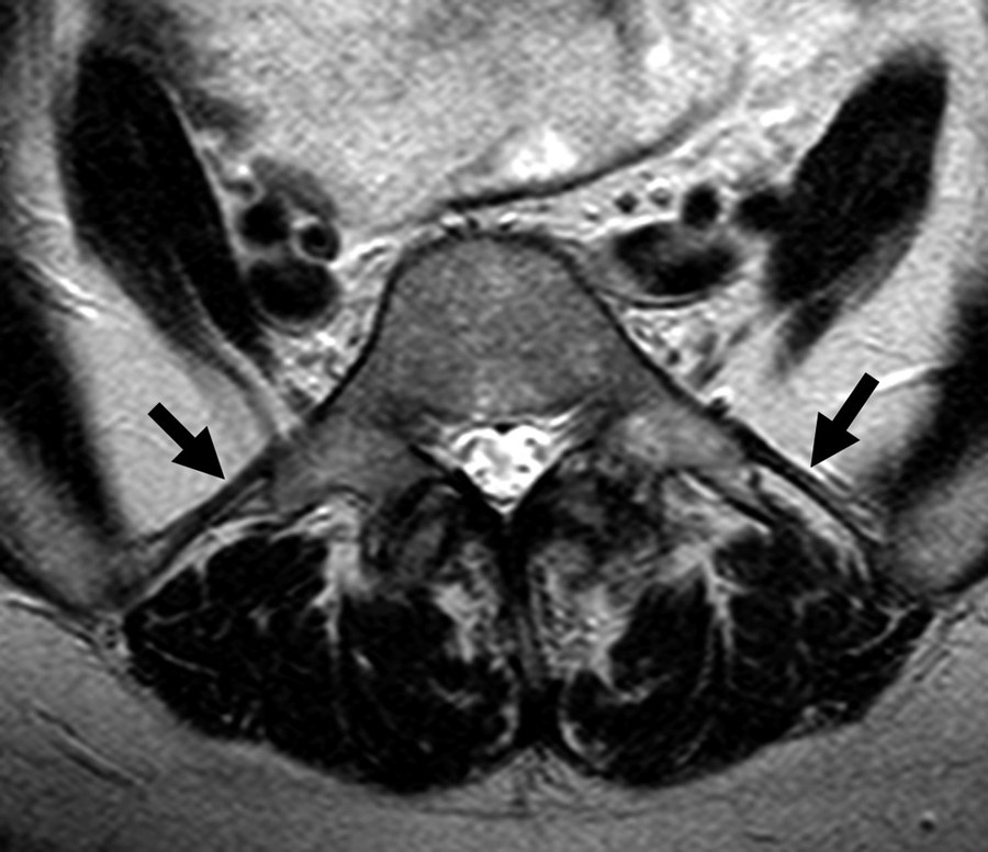
Iliolumbar ligaments (arrows) — low signal intensity on axial T2, running from the L5 transverse processes to the posteromedial iliac crests. Reproduced with attribution from Konin GP, Walz DM. AJNR Am J Neuroradiol 2010;31:1778–86 (Fig 12), open access.
Step 3
Is there a lumbosacral transitional vertebra?
Look for a transverse process at the lowest lumbar-type level that is broadened, articulates with the ala, or is fused to it. The coronal reformat is the plane that shows it; CT characterises it better than anything else, and on radiographs the Ferguson view (AP angled 30° cranially) is the classic projection.
Step 4
Castellvi type
Suffix a = unilateral, b = bilateral. Type I is a dysplastic transverse process at least 19 mm in craniocaudal width with a clear gap; type II articulates; type III is fused.
Drawings reproduced with attribution from Konin GP, Walz DM. AJNR Am J Neuroradiol 2010;31:1778–86 (Fig 2), open access. Classification per Castellvi et al., Spine 1984.
Step 5
Sacralized L5, or lumbarized S1?
Only worth settling if the level drives management. Below are the routes that actually decide it — morphology alone (squaring, wedging) is corroboration, never proof. Every sign below is readable on CT.
Result
Level assignment
Confidence is capped by the imaging tier you chose in step 1.
—
—
confidence
Castellvi classification
Konin & Walz, AJNR 2010 · Fig 2
Type I dysplastic transverse process ≥19 mm craniocaudal with a clear gap to the ala · Type II incomplete transition with a diarthrodial pseudoarticulation between the process and the sacrum · Type III complete osseous fusion of the process to the sacrum · Type IV mixed, type II on one side and type III on the other. Suffix a unilateral, b bilateral (types I–III). Reproduced with attribution from Konin GP, Walz DM. Lumbosacral transitional vertebrae: classification, imaging findings, and clinical relevance. AJNR Am J Neuroradiol 2010;31:1778–86 — doi:10.3174/ajnr.A2036, open access. Classification per Castellvi et al., Spine 1984.
Reference
algorithm, variants, measurements
The gold standard — count from C2, and what each localizer actually buys you
T1 is the first rib-bearing vertebra; L1 is the first non–rib-bearing vertebra below the last ribbed level. Count caudally from C2 on a sagittal whole-spine localizer or scout to the last mobile presacral segment. Normal total is 24 presacral segments (7 cervical, 12 thoracic, 5 lumbar), present in about 91%.
Whole-spine radiographs, CT or MR. The only method that both counts from C2 and distinguishes a hypoplastic 12th rib from an enlarged lumbar transverse process. In practice these are rarely available at the time of reporting.
Sagittal + coronal through the cervicothoracic junction. On CT that means the topogram plus a coronal reformat; on MR, both localizers. Adding the coronal materially increases numbering accuracy, because it is the plane in which dysplastic ribs and lumbar transverse processes separate.
Lower ribs, lumbar spine and sacrum in the field. The everyday body study. You are not counting from C2 at all: you find the last rib-bearing vertebra in the volume and call the next body down L1. That removes every assumption below the ribs, which is why it beats a sagittal topogram — but it cannot exclude an 11- or 13-rib variant or a thoracolumbar transition, because you never see the top of the thoracic spine. Follow each twelfth rib out on the coronal or across consecutive axials; on a single axial image a hypoplastic rib and a hypertrophied transverse process are the same grey blob.
Sagittal alone — CT topogram or MR localizer. Counts from C2 but silently assumes 7 cervical and 12 thoracic vertebrae. It does not account for a thoracolumbar transitional vertebra or an 11- or 13-rib variant, and on MR the large field of view and thick sections often do not resolve enough anatomy to number reliably.
Lumbar coverage alone. Segments are identified correctly in only about 80% of cases with roughly 12% interobserver disagreement. This is the common failure mode on a CT abdomen and pelvis reported without a look at the topogram.
Where an LSTV matters, radiographs remain useful: the Ferguson view (AP angled 30° cranially) profiles the L5 transverse processes and their relationship to the ala. CT gives the best characterisation overall — which is the body reader's advantage, since the coronal reformat that types the transition is already reconstructed. MR is where numbering most often goes wrong, because the cervicothoracic localizer is frequently missing.
Radiographs — and who is actually counting
Count the films yourself. Radiographs already in the record are often not separately reported for numbering, so “correlate with radiographs” is not a disposition on its own — if the images are there, open them and count them. It costs less time than the phone call after a wrong-level injection.
What a lumbar series gives you. The AP usually includes the twelfth ribs, which is all you need to anchor L1 and count down — the same rib-anchored method as the everyday CT, at a fraction of the dose.
The Ferguson view (AP angled 30° cranially) is the classic projection for a transitional segment: it profiles the L5 transverse processes against the sacral ala and separates a wide process with a clear gap from one that articulates.
The lateral shows squaring of a lumbarized upper sacral segment, wedging of a sacralized lowest lumbar body, and the disc height between the transitional segment and the sacrum.
A chest radiograph counts ribs for free — as does the topogram of any prior CT in the record. Either can upgrade a provisional lumbar-only study before you commit to a number.
CT still characterises a transitional segment better than any radiograph. Radiographs are for the count and for the intraoperative correlation, not for typing the transition.
Hypoplastic ribs and riblets — what counts as a rib
The discriminator is a joint, not a length. A rib articulates: there is a costovertebral facet and a lucent cleft between the ossicle and the vertebra. A transverse process is continuous cortex and medullary bone with the neural arch, with no cleft at any window. Follow the structure out on the coronal or across consecutive axials — on one axial image a hypoplastic rib and a hypertrophied transverse process are the same grey blob.
Farshad-Amacker's scheme makes that operational at the lowest rib-bearing segment:
Short (hypoplastic) rib — under 3.8 cm, but its head still reaches a vertebral facet. The segment is thoracic. A riblet is still a rib.
Accessory ossification centre (a true lumbar rib) — does not reach a vertebral facet and has no true joint with the costal process. The segment is lumbar.
Mixed — a separated ossicle meeting neither definition. Describe it and state what you assumed; do not silently pick one.
Check both sides before you commit. Lumbar ribs are bilateral in about two-thirds of cases, so a solitary unilateral ossicle deserves a harder look at the contralateral side. Pooled prevalence of lumbar ribs is about 2.1% in the general population and 1.6% among asymptomatic people; thoracolumbar transitional vertebrae run about 4.1%, and an anomalous total vertebral count about 8.2%.
The two strategies disagree, and that is the whole problem. Counting caudally from C2 while assuming 12 thoracic vertebrae, and identifying the last rib-bearing segment morphologically, give different numbers in exactly these patients. Neither is the anointed method, so the report has to say which one you used — a bare level name is not reconstructable by anyone downstream.
The association that should change your behaviour: an LSTV is significantly associated with an anomalous total vertebral count. Finding a transitional segment at the bottom of the spine is a reason to go and check the top, not a reason to settle.
How to write it when the junction is ambiguous. An explicit formula beats a bare level — C7–T12–TLTV–L5–LSTV — or name the absolute count ("the 19th vertebral body"). Either lets the next reader reconstruct your numbering even if they use the other convention.
Numeric variants and the blind spot
Lumbar-type count: 4 (~2.6%), 5 (~89%), 6 (~8.2%). Roughly 2.6% of people have 23 and ~3.3% have 25 presacral segments, and a thoracolumbar transitional vertebra or rib variant (11 or 13 ribs) can shift the count independently — so do not assume 12 rib pairs.
The blind spot: about 2.6% of four-lumbar and ~2.9% of six-lumbar spines have no transitional vertebra and look exactly like a modal L5 spine on lumbar imaging. These numeric-only variants are undetectable without whole-spine correlation — though under our convention the count itself is what licenses the words. Six lumbar-type bodies means the uppermost sacral segment is lumbar-type, which is what “lumbarized S1” says; four means the lowest lumbar segment is sacral-type. Castellvi typing is a separate axis, describing the transverse process, so “no transitional vertebra” on that axis does not stop you calling a six-lumbar spine a lumbarized S1.
Fallback landmarks when only lumbar imaging is available
Ranked by reliability; each is imperfect, and none makes the number definitive.
Psoas proximal insertion: the most cephalad vertebra with psoas covering more than half the body — on coronal CT reformats, or coronal T2/STIR on MR (98.5% modal, 81.4% when an LSTV is present). If psoas does not insert five bodies above the apparent lumbosacral joint, a numeric variant is likely — an automatic trigger to request whole-spine imaging.
Iliolumbar ligament (axial): a single or double band running from the transverse process to the posteromedial iliac crest — low signal on axial T1 or T2, and on CT a soft-tissue band identifiable on soft-tissue windows, though less conspicuous than on MR. It is identifiable in about 86% of people and arises from L5 in about 97% of those — so it identifies a segment rather than counting one: if no iliolumbar ligament arises from the level above the transitional vertebra, call the transitional vertebra L5; if a ligament does arise above it, that segment is L5 and the transitional vertebra is S1. The rule assumes 7 cervical, 12 thoracic and 5 lumbar vertebrae, so it fails in the presence of segmentation anomalies or a thoracolumbar transition — exactly the situation in which you most want it. Ligaments above an LSTV are also thinner and weaker, which can make them harder to see.
Iliac crest tangent sign (coronal, CT reformat or MR): sensitivity ~81%, specificity 64–88%, interreader κ ~0.75. Usable without advanced technique, but not definitive.
Do NOT number off the aortic bifurcation, right renal artery, SMA or conus. The right renal artery is usually at the L1–2 disc but is out of field or at another level 25% of the time; the aortic bifurcation is at L4 in only 83%; the conus is too variable to be used as a landmark at all.
Sacralized L5 vs lumbarized S1 — what actually decides it
Quantitative CT (mid-sagittal) outperforms morphology when whole-spine imaging is unavailable:
Anterior-edge vertebral angle (AVA) of the lowest lumbar-type vertebra: cutoff 73.0° (accuracy 91.1%).
RISE ratio (inferior/superior endplate length of the uppermost sacral-type vertebra): cutoff 0.79 (accuracy 90.3%).
A high iliac crest line plus a posterior angle <20° identified lumbarization in 95.8% of cases.
Corroborating morphology on MR — supportive, never sufficient on its own:
Squaring vs wedging. A lumbarized S1 looks squared on the sagittal; a sacralized L5 looks wedged. The quantitative version is the ratio of AP diameter of the superior to inferior endplate, ≤1.37 for "squared" — but squaring and wedging are a spectrum and cannot definitively identify an LSTV.
The S1–S2 disc. O'Driscoll graded sagittal S1–2 disc morphology in four types: 1 no disc material, 2 a small residual disc shorter than the sacrum (both typical of non-transitional spines), 3 a well-formed disc spanning the full AP length of the sacrum, 4 type 3 plus squaring of the presumed upper sacral segment. Type 4 correlates well with a Castellvi III or IV S1 LSTV. The system is sagittal and disc-based, so it misses Castellvi type II and some type III — check axial or coronal images before relying on it.
Facet joints. With a sacralized L5 the L5–S1 facets are hypoplastic or absent when the process is fully fused. With a lumbarized S1 there are often well-formed facet joints at S1–S2, where fusion is the norm.
Disc height. The disc between a lumbar transitional segment and the sacrum is lower than a normal L5–S1 disc; conversely a lumbarized S1 has a larger-than-vestigial S1–S2 disc.
Combined nomenclature & clinical stakes
DHMC convention: the lumbar spine is always L1–L5. We do not label a lowest mobile presacral segment L4 or L6. A four-lumbar-type spine is a sacralized L5; a six-lumbar-type spine is a lumbarized S1. Because of this the count of lumbar-type bodies settles sacralization versus lumbarization on its own — which is why the tool stops asking once you have counted. Report the designation and the morphology together, e.g. L5 IIa or S1 IIIb, and state (a) how many lumbar-type bodies there are, (b) the numeric variant, (c) the Castellvi type, and (d) the confidence source. Some of the literature (and outside reports) number these L4 and L6 instead — say the count so the two can be reconciled.
Why it matters: most wrong-level spine surgery happens in patients with variant anatomy, and errors cluster where a lumbar study was reported without accompanying radiographs, a topogram or a cervicothoracic localizer. Miscounting occurred in 54% of LSTV cases in one surgical series and altered the operative plan in 15%. Because the surgeon confirms level on intraoperative radiographs, the preoperative CT or MR must be explicitly correlated with them — say the number, say how you arrived at it, and say what would change it.
One clinical caveat worth a sentence in the report: the functional L5 nerve root always originates at the lowest mobile lumbosacral level. With a sacralized L5 the L4 root does the usual work of L5; with a lumbarized S1 the S1 root does. That is why radicular findings can appear one level off from the anatomy.
Sources
Konin GP, Walz DM. Lumbosacral transitional vertebrae: classification, imaging findings, and clinical relevance. AJNR Am J Neuroradiol 2010;31:1778–86 — doi:10.3174/ajnr.A2036 (open access; Figs 2 and 12 reproduced with attribution). · Castellvi AE, Goldstein LA, Chan DPK. Lumbosacral transitional vertebrae and their relationship with lumbar extradural defects. Spine 1984. · Hughes RJ, Saifuddin A. Numbering of lumbosacral transitional vertebrae on MRI. AJR 2006. · Peh WCG et al. Determining the lumbar vertebral segments on MRI. Spine 1999. · Hahn PY et al. Cervicothoracic MR localizer for vertebral enumeration. · O'Driscoll CM et al. Sagittal MR classification of S1–2 disc morphology. · Wigh RE, Anthony HF. Transitional lumbosacral discs. · Sabri SA, Laynes RA, Hoffman CJ et al. Implications of lumbosacral transitional anatomy on vertebral numbering (3147 full-length spine radiographs). Neurosurg Focus 2025. · Paik NC, Lim CS, Jang HS. Numeric and morphological verification of lumbosacral segments in 8280 consecutive patients. Spine 2013. · Lumbosacral transitional vertebra in adolescents. Biomed Res Int 2021. · Carrino JA et al. Effect of spinal segment variants on numbering vertebral levels at lumbar MR imaging. Radiology 2011. · Farshad-Amacker NA et al., morphologic classification of the lowest rib-bearing segment; see also Spinal enumeration by morphologic analysis of spinal variants. Korean J Radiol 2018. · Prevalence and characteristics of lumbar ribs: a meta-analysis. Surg Radiol Anat 2024. Text paraphrased and summarised in our own words; figures reproduced with attribution and linked to the originals.
 Back to Menu